Study of the radiographic parameters of normal ankles
literature review and technical recommendations
DOI:
https://doi.org/10.30795/jfootankle.2020.v14.1125Keywords:
Ankle, Ankle joint, X-rays, Radiography, ArthritisAbstract
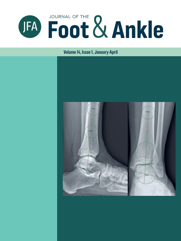
Objective: The authors carried out a bibliographic search for the radiographic parameters used to determine tibiotalar joint alignment, and suggest a set of parameters that constitute the minimum radiographic evaluation sufficient for the proper assessment of tibiotalar alignment. Methods: The search was conducted between May 2019 and January 2020 on the online platforms PudMed and Google Scholar with the following terms, used separately or jointly: “ankle arthritis, radiographic measurement, ankle alignment, alignment, anterior ankle instability, X-ray, and ankle injury”. Results: We selected twelve studies evaluating radiographic patterns of normal ankles, and identified a total of 15 radiographic measurements. Conclusion: The authors believe that a minimum radiographic assessment of tibiotalar alignment should include the following parameters on the anteroposterior radiograph: the distal tibial articular angle, the talar tilt and talus center migration. On the lateral radiograph, it should include: lateral distal tibial angle and lateral talar station. Level of Evidence V; Diagnostic Study; Expert Opinion.